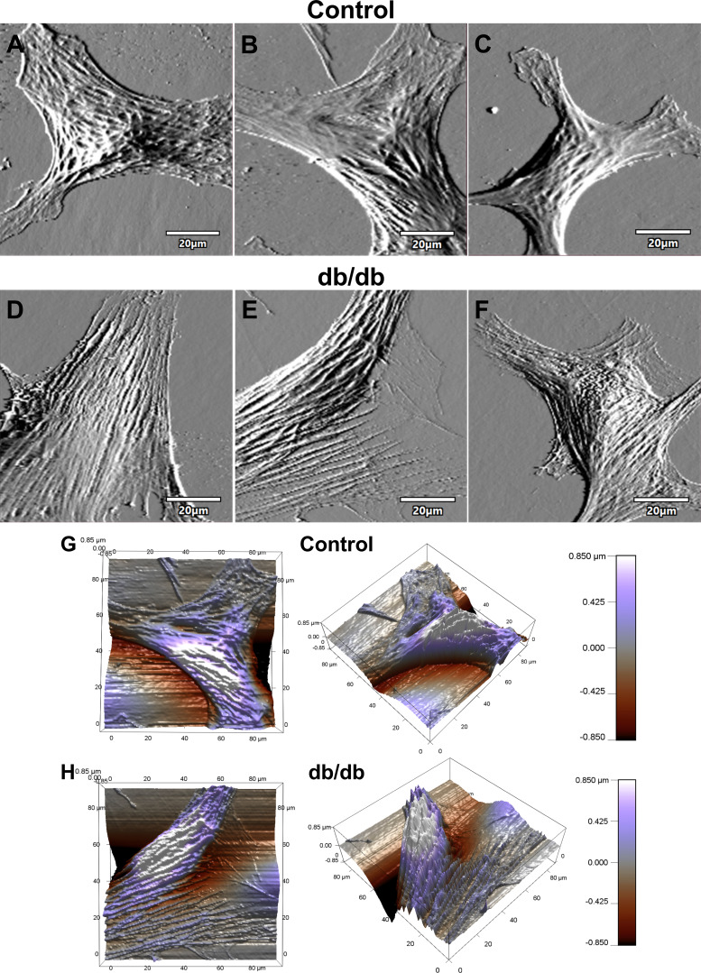
Fig. 3.
Coronary vascular smooth muscle cell (VSMC) topography. A–F: representative contact-mode images of coronary VSMCs from control and diabetic mice: deflection images of normal control coronary VSMC (A–C) and diabetic coronary VSMC (D–F). G and H: representative 3-dimensional z-sensor images of normal control VSMC (G) and diabetic VSMC (H).

