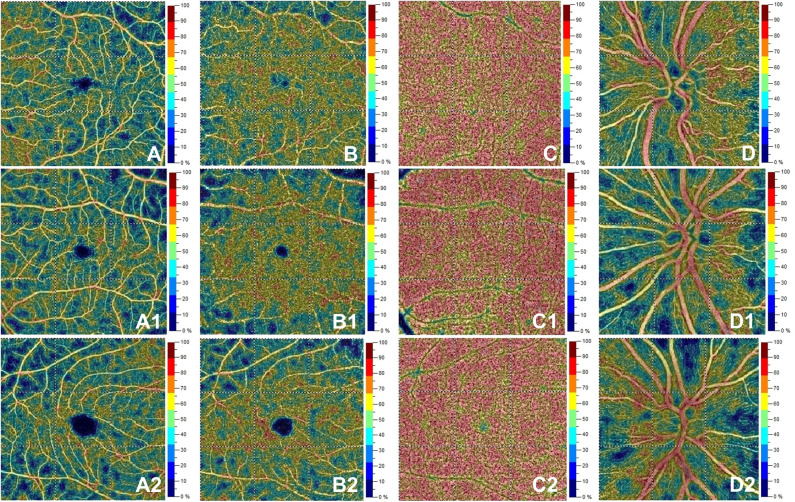
Figure 2.
Optical coherence tomography angiography (OCT-A) images from a healthy subject' s left eye (male, 28 years) in the first row show normal vessel density in superficial capillary plexus (A), deep capillary plexus (B), choriocapillaris (C) and radial peripapillary capillary plexus (D). The second row depicts OCTA features in the left eye for a patient (female, 28 years) with initial demyelinating event. OCTA reveals normal vessel density in superficial capillary plexus (A1), deep capillary plexus (B1), choriocapillaris (C1) with a decrease for vessel density in the radial peripapillary capillary plexus (D1). The bottom row shows a patient's right eye (male, 29 years) affected by relapsing-remitting multiple sclerosis. Here, vessel density is reduced in the superficial capillary plexus (A2) and radial peripapillary capillary plexus (D2) without vessel density changes in the deep capillary plexus (B2) and choriocapillaris (C2).

