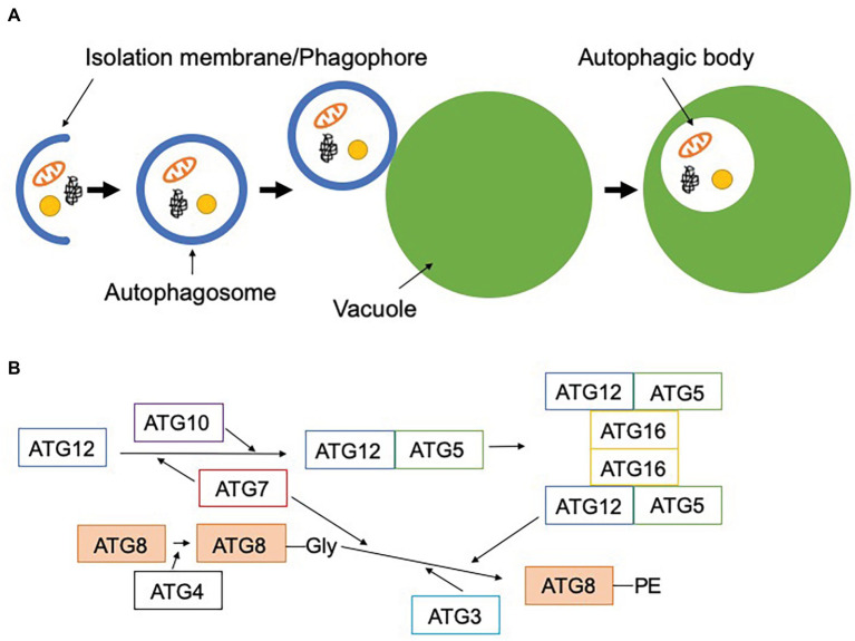
Figure 1.
Scheme of macroautophagy. (A) Macroautophagy starts with the formation of the isolation membrane (phagophore) in the cytosol. This engulfs cytoplasmic components and forms the double membrane-bound autophagosome. The outer membrane of the autophagosome fuses with the vacuolar membrane to release a single membrane-bound autophagic body into the vacuole. (B) Two ubiquitin-like conjugation systems are involved in the lipidation of ATG8. First, ATG12 is conjugated to ATG5 by ATG7 (E1-like) and ATG10 (E2-like), and ATG12-ATG5 forms a complex with ATG16. ATG8 is cleaved by ATG4, resulting in the exposure of glycine at its carboxyl terminus. This processed ATG8 is conjugated to phosphatidylethanolamine by ATG7 (E1-like), ATG3 (E2-like), and the dimeric ATG12-ATG5-ATG16 complex (E3-like). Lipidated ATG8 can be localized to the autophagosomal membrane.

