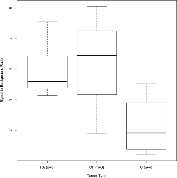
FIGURE 3.

NIR tumor fluorescence varies based on tumor type. All 8 pituitary adenomas demonstrated NIR positivity and fluoresced with an average SBR of 3.9 ± 0.8. Craniopharyngiomas (CP) had the highest average SBR of 4.1 ± 1.7, which was not statistically different from the average SBR of pituitary adenomas (PA; Wilcoxon rank sum, P = .78). However, the average SBRs of both pituitary adenomas and cranoipharyngiomas were statistically distinct from that of chordomas (C; SBR = 2.1 ± 0.6).
