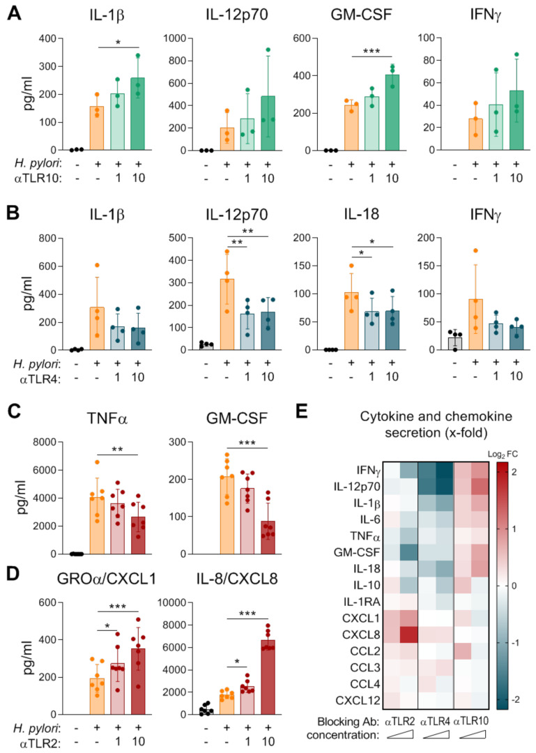
Figure 2.
Impacts of TLR2, TLR4, and TLR10 on cytokine and chemokine secretion by cDC2s in response to H. pylori. cDC2s were infected with H. pylori P12 (MOI 5). Twenty minutes prior to infection, the cDC2s were treated with blocking antibodies to (A) TLR10, (B) TLR4, or (C,D) TLR2 at a concentration of 1 µg/mL or 10 µg/mL. Cytokine and chemokine secretion was monitored 16 h post H. pylori infection by multiplex assay. Dots represent individual donors, bars and lines show means ± SDs. For statistical analysis, repeated-measures ANOVA with Dunnett’s post-hoc test was performed. (* p ≤ 0.05, ** p ≤ 0.01, *** p ≤ 0.001). (E) Log2 fold change (Log2 FC) was calculated using log2 of the following ratio, mean of inhibitor-treated samples:mean of H. pylori-infected samples. Red: up-regulation, blue: down-regulation.

