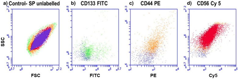
Figure 1.
Fluorescence protein detection, using flow cytometry. (a) Control SP of cells that are unlabeled. (b) Lung CSCs positive for CD133 (FITC), (c) lung CSCs positive for CD44 (PE) and (d) lung CSCs positive for CD56 (Cy5). All positive samples are overlaid with the control, to distinguish between the color shifts.

