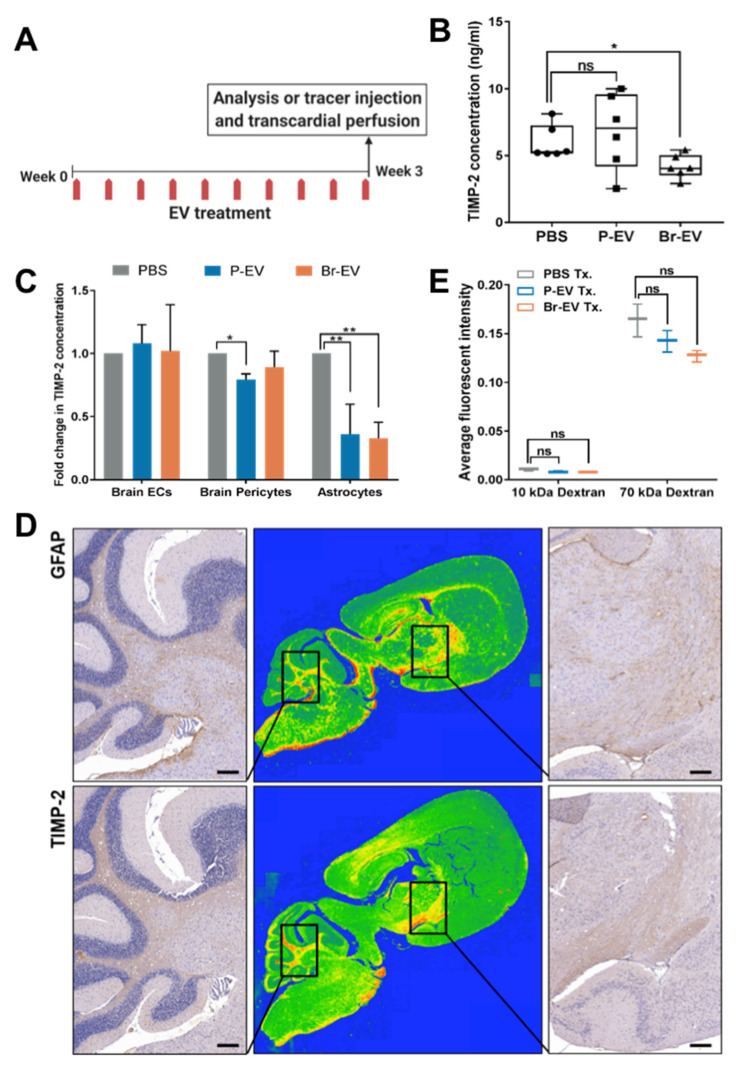
Figure 3.
Br-EVs decrease the astrocyte expression of the tissue inhibitor of matrix metalloproteinases-2 (TIMP-2). (A) Schematic showing the EV functional study design. (B) Average concentration of TIMP-2 in brain tissue homogenates measured by a mouse TIMP-2 enzyme-linked immunosorbent assay (ELISA) (mean ± SD; n = six mice per group). Statistical analysis was performed using the Mann–Whitney test. (C) Average fold change in concentration of TIMP-2 in conditioned media of brain endothelial cells, pericytes and astrocytes treated with PBS, P-, and Br-EVs (mean ± SD; three independent experiments). Statistical analysis was performed using two-way ANOVA with Sidak’s multiple comparison tests. (D) Representative images of mouse brain sections immunostained with anti-GFAP (upper panels) and anti-TIMP-2 (lower panels), demonstrating colocalization of GFAP astrocyte marker and TIMP-2. Middle panels represent a colormap of areas of protein enrichment (three independent experiments). Scale bar, 200 µm. (E) Average fluorescence intensity in perfused brain tissue homogenates collected 45 min following injection of a combination of 10 KDa Alexa647 dextran and 70 KDa FITC dextran (mean ± SD; n = three mice per group). Statistical analysis was performed using the Mann–Whitney test. In all panels: ns, not significant; * p ≤ 0.05; ** p ≤ 0.01.

