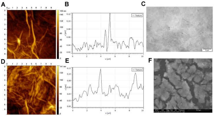
Figure 1.
Graphene oxide scaffold morphology. (A) Atomic force microscopy images and (B) a topography model of the surface of the graphene oxide scaffold on a polystyrene culture plate. (C) Transmission electron microscopy image of graphene oxide. (D) Atomic force microscopy images and (E) a topography model of the graphene oxide scaffold surface on a flat silicon wafer. (F) Scanning electron microscopy image of the graphene oxide scaffold.

