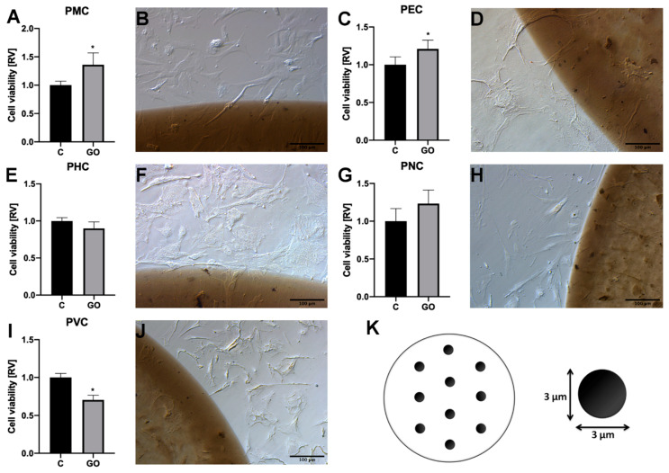
Figure 2.
Analysis of the cell localization and viability on the GO scaffold. (A) Viability and (B) morphologies of progenitor muscle cells from hind limb (PMC) on the GO scaffold. (C) Viability and (D) morphology of progenitor eye cells (PEC) on the GO scaffold. (E) Viability and (F) morphology of progenitor heart-derived cells (PHC) on the GO scaffold. (G) Viability and (H) morphology of cells derived from brain (progenitor nerve cells, PNC) on the GO scaffold. (I) Viability and (J) morphology of cells from a chorioallantoic membrane’s blood vessel (progenitor vessel cells, PVC) on the GO scaffold. Morphology was assessed on the edge of the GO scaffold via light microscopy with phase contrast and 200 × magnification. Statistical significance is indicated with different superscripts (unpaired t-test; p < 0.05). (K) Pattern of GO islands used in cell localization and morphology analysis. Acronyms: C, control; GO, graphene oxide; RV, relative value.

