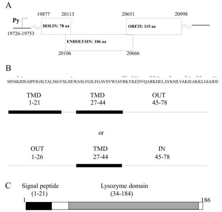
Figure 1.
In silico analysis of HP1 phage lytic locus and protein proprieties. (A) Organization of lysis locus within the genome of HP1 phage. Numbers indicate positions within the genome. (B) Amino acid sequence showing charged amino acids of holin protein. Two potential organizations of holin within the cell membrane according to SOSUI (2 trans membrane domains-TMD) and other algorithms (1TMD). (C) HP1 phage endolysin domain organization.

