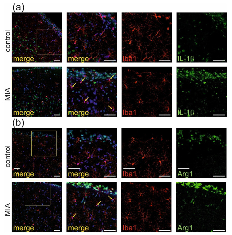
Figure 6.
MIA induces activation of microglia a in brain cortex of adolescent male rats. Immunohistochemical analysis of somatosensory cortex illustrating microglia cells (Iba-1—red) in control and MIA-exposed groups. Co-expression of activation markers, (a) IL-1β (green) and (b) arginase-1 (green), with Iba1-positive cells has been observed. The nuclei were counterstained with DAPI (blue). Scale bar = 50 μm. Yellow arrows indicate the cells which display the most evident co-localisation.

