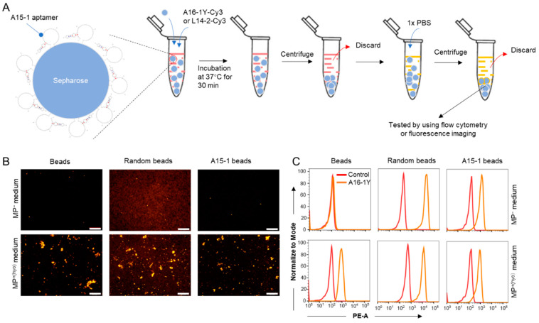
Figure 8.
Detection of mycoplasma-contaminated culture medium. Streptavidin-immobilized sepharose and the A16-1Y aptamer were used to distinguish between MP+ and MP− cell culture media. (A) Flow chart of the experiments for mycoplasma detection in cell culture medium. Indicator beads were incubated with MP− or MP+ cell culture medium containing [100 nM] Cy3-labeled A16-1Y or control aptamer at 37 °C for 30 min. Then, the fluorescence of those aptamer-bound beads was observed using a fluorescent microscope or a flow cytometer. Red in the tube indicates cell culture medium, yellow in the tube indicates 1 × PBS. (B) Fluorescence microscopy observation of the Cy3-labeled A16-1Y aptamer binding with M. hyorhinis-adherent sepharose. (C) Histogram plots of flow cytometric data representing Cy3-labeled A16-1Y aptamer binding with M. hyorhinis-adherent sepharose. Scale bar = 500 μm.

