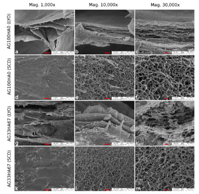
Figure 3.
SEM images of agarose lyophilized (LYO) (a–c) and supercritically-dried (SCD) (d–f) and agarose/hydroxyapatite (33/76 w%) composite LYO (g–i) and SCD (k–m) at three different magnifications. The scale bars are 10 μm (left), 1 μm (middle), and 0.2 μm (right), respectively. Reproduced from Witzler et al., 2019 [171]. Open Access Copyright Permission (Creative Commons CC BY license).

