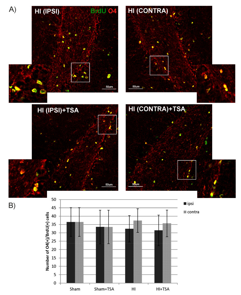
Figure 7.
TSA does not affect the number of non-myelinating oligodendrocytes in DG after HI. (A) The confocal photomicrographs show double-labeled (BrdU/O4-positive) cells in DG of HI animals D28 with or without TSA treatment. Enlargements present areas marked in rectangles. Scale bar 50 µm. (B) The graph shows the number of BrdU/O4 labeled cells quantified in the DG area (0.36 mm2). Values represent mean ± SD of five animals per experimental group. Two-way ANOVA tests did not indicate significant differences in the number of BrdU/O4-positive cells between the investigated groups. Abbreviations: ipsi—ipsilateral, contra—contralateral.

