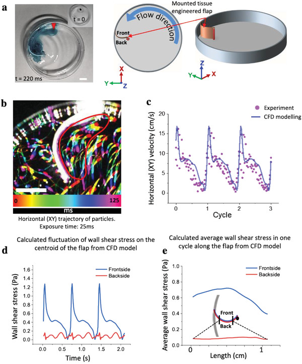
Figure 1.

Mounting of an engineered tissue scaffold in the orbital shaker dish platform together with the CFD analysis of the oscillatory traveling wave. a) Actual image of the petri dish with a drop of ink (image inset, red arrow) to illustrate the fluid motion (Movie S4, Supporting Information), and two schemes to show how the rectangular tissue scaffold made from biodegradable PGA‐P4HB is mounted in the dish, together with defining the three coordinates used in later figures. The tissue scaffold was attached with one end to the dish wall and with the other end to an upright post, located 1 cm away from the wall, thereby creating a convexly curved barrier facing the flow direction. b) The horizontal (XY) trajectories of particles (compiled sequence of five images sequentially colored in rainbow colors) were visualized by short exposure (25 ms) with the aid of a high‐speed camera. The lengths of the experimental particle trajectories were used to calculate the corresponding velocities in the encircled region (as marked by the red line). Scale bars 5 mm. c) To validate the CFD calculations, the time‐resolved experimentally calculated horizontal velocities (dots) during three cycles in front of the tissue are shown to be in good agreement with the average flow velocities as obtained by CFD. d) Fluctuations of shear stresses on one spot at centroid of flap and e) average of wall shear stress along a horizontal curve on the frontside (blue line) and backside (red line) of flap in a cycle.
