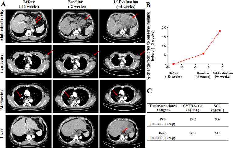
Figure 1.
Case study of a patient in his mid-40s with HPD during immunotherapy. (A) CT scans were performed 13 weeks before starting anti-PD-1 treatment (column 1), at baseline (2 weeks before starting immunotherapy, column 2), and at first evaluation (4 weeks after starting immunotherapy, column 3). CT scans from lines 1 to 4 revealed the changes in lymph nodes in the abdominal cavity, left axilla and mediastina, respectively. New liver lesion appeared. The red arrows indicate tumer lesions.(B) Rate of change in growth pattern in the patient, who developed HPD to camrelizumab. Compared with the tumor image (−13 weeks), the tumor lesions at baseline (−2 weeks) and at first evaluation (4 weeks after starting immunotherapy) showed approximately 57% and 181% increases (79% increase compared with baseline imaging), respectively; 2.5-fold increase in progressive pace compared with preimmunotherapy. (C) Changes in tumor-associated antigens before and after immunotherapy. CYFRA21-1, cytokeratin-19 fragment; HPD, hyperprogressive disease; PD-1, programmed cell death 1; SCC, squamous cell carcinoma.

