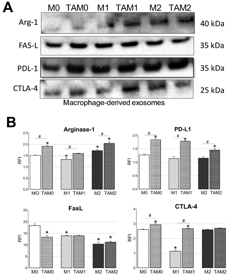
Figure 3.
Immunosuppressive cargos of exosomes produced by macrophages or TAMs. (A) Representative Western blots of exosomes isolated from macrophages or TAMs. Equal amounts of exosomal protein (10 μg) were loaded per lane; (B) Flow cytometry results for the detection of PDL-1, FasL, CTLA-4, and Arginase-1 carried on exosomes produced by macrophages or TAMs. Exosomes were immunocaptured with anti-CD63 mAb for on-bead flow cytometry as described in Materials and Methods. Data are relative fluorescence intensity (RFI) values ± SEM from three independent experiments Data were analyzed by ANOVA followed by Tukey post hoc. *Significantly different from the control at p < 0.05 and # Significant difference between macrophages and TAMs at p < 0.05.

