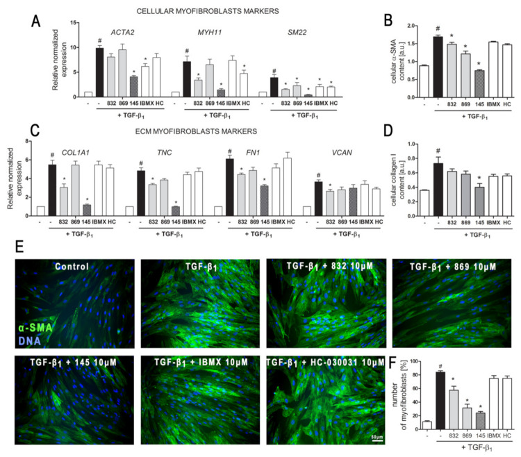
Figure 3.
Compound 145 significantly reduced TGF-β1-induced MRC-5 transition into myofibroblasts. MRC-5 were pre-incubated for 1 h with the studied compounds (10 µM) and then cultured for 24 h (A, C) or 48 h (B, D–F) in TGF-β1 (5 ng/mL). (A, C) qPCR was carried out to analyze ACTA2, MYH11, SM22, COL1A1, TNC, FN1, and VCAN transcript levels. Samples were run three times in duplicates. Cellular α-smooth muscle actin (α-SMA) (B) and collagen I (D) protein level was determined by in-cell ELISA; n = 8. (E) To visualize α-SMA positive stress fibers, MRC-5 were fixed, permeabilized, blocked, and labeled with anti-α-SMA antibody, followed by Alexa Fluor 488 conjugated antibody, nuclei were stained with Hoechst 33342 dye. Microphotographs were taken using a Leica DMiL LED Fluo microscope, 40× objective, bar = 50 µm. (F) Myofibroblasts (MRC-5 positive for α-SMA) were counted in 20 randomly selected fields of view and expressed as a percentage of the entire MRC-5 population. Each bar represents the mean value (± S.E.M.). The results were considered statistically significant at the p level of 0.05, against control (#) and TGF-β1 (*).

