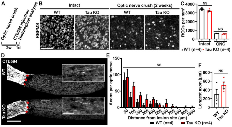
Figure 2.
Tau deletion does not influence retinal survival and axonal growth. (A). Experimental timeline. Optic nerve crush (ONC) was performed on the left eye of WT and Tau KO mice. Two weeks later, axons were traced with CTb594 the day before animal sacrifice. (B). Influence of Tau expression on the survival of retinal ganglion cells (RGCs) 2 weeks after ONC in WT and Tau KO mice. Flat-mounted retinae were immunostained for RNA-Binding Protein with Multiple Splicing (RBPMS) to determine RGC density. Scale bar: 50 µm. (C). Quantitative analysis of RGC survival did not show difference between WT and Tau KO mice (n = 4 mice per group). Data are expressed as mean/mm2 ± S.E.M. Statistics: Student’s t-test. NS: Non-significant. (D). Influence of Tau deletion on axonal outgrowth at 2 weeks ONC. The growth of injured RGC axons was examined after the lesion site (red stars) in optic nerve cryosections of WT and Tau KO mice. Close-ups showed axonal sprouts beyond the injury site. Scale bars: 250 µm, close-up: 50 µm. (E). Quantitative analysis of axonal outgrowth. The number of axons per optic nerve was estimated in injured WT and Tau KO animals at distances ranging from 50 to 1000 μm past the injury site (n = 4 mice per group). Despite a trend toward an increase, Tau KO optic nerves did not display more growing axons than those of WT mice. Data are expressed as mean ± S.E.M. Statistics: Student’s t-test. NS: Non-significant. (F). Measurement of the longest axons in injured optic nerves. The longest axons measured in lesioned optic nerves did not reveal longer maximal extensions between Tau KO and WT mice (n = 4 mice per group). Data are expressed as mean ± S.E.M. Statistics: Student’s t-test. NS: Non-significant.

