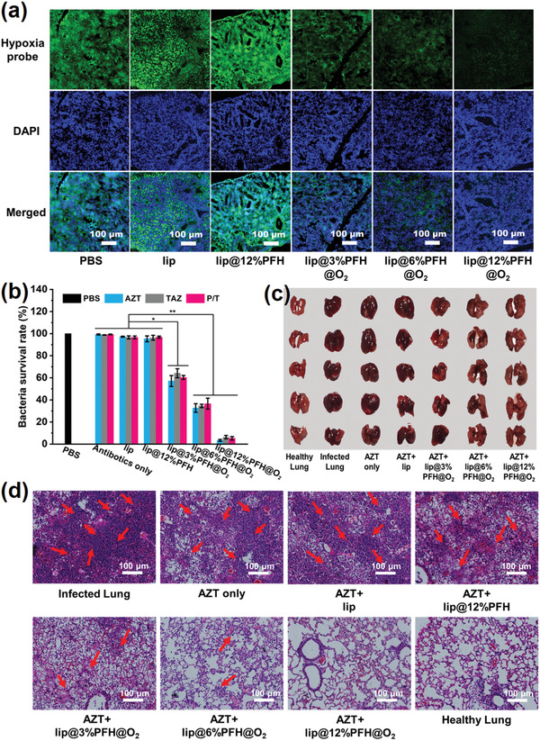
Figure 4.

a) Representative immunofluorescence images of lungs infected by P. aeruginosa biofilm with different treatments stained by the hypoxyprobe. The nuclei and hypoxia areas are stained with DAPI (blue) and anti‐pimonidazole antibody (green), respectively. Scale bar: 100 µm; b) Related quantitative results of standard plate counting assay after different treatments with same antibiotic concentration (1 mg mL−1) in the P. aeruginosa biofilm infected chronic pneumonia model; c) Digital photographs of P. aeruginosa biofilm infected lungs treated with different nanoparticles with same aztreonam concentration (AZT, 1 mg mL−1) on 2nd day; d) Histological photographs of infected lungs treated with different nanoparticles with same aztreonam concentration (AZT, 1 mg mL−1) on 2nd day, the inflammatory cells marked by red arrows.
