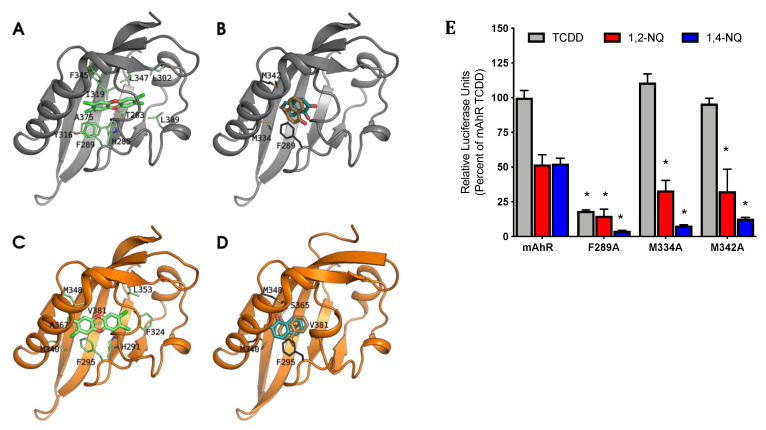
Figure 6.
NQ interact with specific residues within the AhR binding cavity that are required for AhR functional activity. The models are shown as cartoons (mAhR in gray, hAhR in orange), and TCDD and NQs as sticks. The residues that mainly contribute to ΔGbind are also shown as sticks (green for TCDD and dark gray for NQs). (A) TCDD pose in the mAhR LBD, taken as reference from previous analyses [44,45,46]: (B) NQs bind differently in the mAhR LBD compared to TCDD: the main interactions are with M334, M342, and F289. (C) TCDD pose in the hAhR: due to the steric hindrance of V381 both the position and the laying plane of the molecule within the cavity are different compared to the pose in mAhR. (D) The NQs poses in the hAhR are in the same relative position in the binding site as in the mAhR LBD, but additional stabilization provided by ligand interactions with V381 and S365 result in differences in NQ orientations. (E) COS-1 cells transiently transfected with wild-type or mutant mAhR pGudLuc6.1 were incubated with DMSO (0.1%, v/v), TCDD (10 nM), 1,2-NQ (5 μM), or 1,4-NQ (5 μM) for 18 to 22 h. Luciferase activity was measured, corrected for background activity (DMSO), and normalized to TCDD activity induced in mAhR transfected cells. Values represent the mean ± standard deviation of three independent experiments. Asterisks (*) indicate those values that are significantly reduced relative to that of mAhR at p < 0.05 as determined by Two-Way ANOVA.

