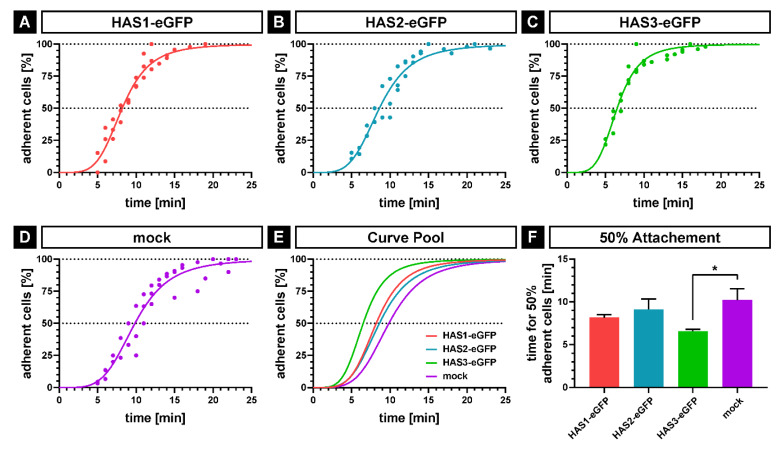
Figure 3.
Analysis of initial cell attachment by time-lapse microscopy. SCP1-HAS-eGFP and SCP1-mock cells were incubated for 48 h in culture medium in the presence of 10 mM GlcNAc including the last 24 h under serum-free conditions. The cells were detached by accutase treatment and were seeded onto uncoated tissue culture polystyrene dishes. Cell attachment was determined by the formation of the first protrusion and the whole process was imaged with 60 frames/h. (A–D) Nonlinear regression by sigmoidal four-parameter-logistic; dots indicate the adhesion of a minimum of one cell at the corresponding time point, showing values of three independent experiments. (E) Overlay of the nonlinear regression curves of the four cell lines. (F) Mean values for 50% adherent cells calculated by sigmoidal four-parameter-logistic for each of the three independent experiments. Error bars represent SD, the asterisk indicates a p-Value < 0.05 (*).

