Figure 3.
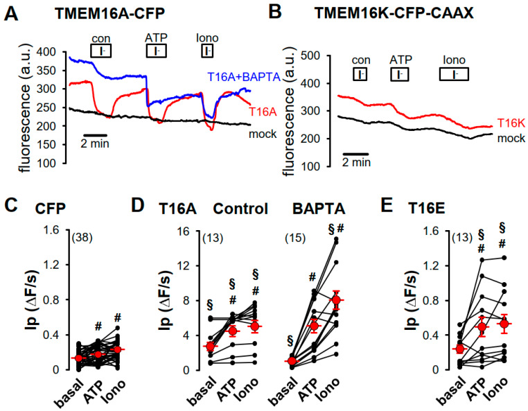
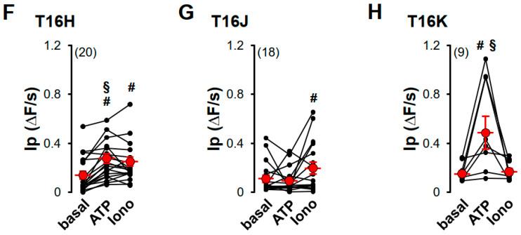
Purinergic stimulation and increase of intracellular Ca2+ concentration by ionomycin induced iodide permeability in HEK293-YFP cells expressing plasma membrane-targeted TMEM16 proteins. (A) Original recording of TMEM16A-CFP-expressing HEK293-YFP cells. Application of 20 mM iodide under control (con) in the presence of 100 µM ATP (ATP) or in the presence of 1 µM ionomycin (Iono) caused in TMEM16A-CFP-expressing HEK293-YFP cells (red, T16A) a strong but in TMEM16A-CFP-non-expressing HEK293-YFP cells (black, mock) only a weak transient quenching of the YFP signal, indicating enhanced iodide permeability through TMEM16A-CFP expression. The Ca2+-chelator BAPTA reduced iodide quenching under control conditions in TMEM16A-CFP-expressing HEK293-YFP cells (blue, T16A + BAPTA). (B) Original recording of TMEM16K-CFP-CAAX-expressing HEK293-YFP cells (red, T16K). Twenty mM iodide induced a slight increase of the YFP-quenching under control (con) and in the presence of 1 µM ionomycin (Iono), like in TMEM16K-CFP-CAAX-non-expressing HEK293-YFP cells (black, mock) but significantly increased YFP-quenching in the presence of 100 µM ATP when compared to control cells (mock). Summaries of iodide permeability (IP) measured by initial slope of fluorescence decrease (ΔF/s) of (C) CFP-expressing cells under basal condition (basal), in the presence of 100 µM ATP (ATP) or in the presence of 1 µM ionomycin (Iono); (D) of TMEM16A-CFP (T16A)-expressing HEK293-YFP cells under control condition or pre-incubated with 50 µM BAPTA for 30 min at RT; (E) TMEM16E-CFP-CAAX (T16E)-, (F) TMEM16H-CFP-CAAX (T16H)-, (G) TMEM16J-CFP-CAAX (T16J)-, or (H) TMEM16K-CFP-CAAX (T16K)-expressing HEK293-YFP cells. (Number of cells measured), # paired t-test, α < 0.05, § unpaired t-test to control and CFP, resepectively, α < 0.05, and analysis of variance (ANOVA) to CFP, α < 0.05.


