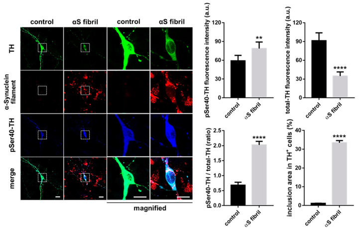
Figure 4.
Exposure to α-Synuclein fibrils formed intracellular filamentous inclusions, which was accompanied by the acceleration of TH phosphorylation and the reduction of total TH protein in the cultured dopaminergic neurons in the presence of cycloheximide. Scale bars indicate 10 μm. The right columns show the quantified data (Student’s t-test, **** p < 0.0001, ** p < 0.01, n > 20). αS indicate α-Synuclein.

