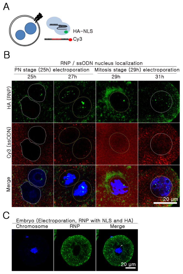
Figure 2.
Nuclear localization of RNP and ssODN. (A) Experimental design. (B) RNP (HA-conjugated SpCas9 and sgRNAs targeting Rosa26 locus) and Cy-3-conjugated 100 bp-sized ssODN were electroporated into embryos at 25 h and 29 h after hCG injection. Next, half were immunostained with HA-Alexa 488 (green) mAb, and the other half were stained after two hours with the same target. Representative images are shown. The related videos are presented in Videos 1 and 2, Supplementary Materials. White solid line: intact nuclear membrane; white dotted line: disappeared nuclear membrane. (C) Physical barriers outside the chromosome (white arrow); blue: chromosome; red: ssODN; green: RNP.

