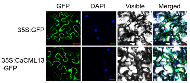
Figure 3.
Subcellular localization of CaCML13 in N. benthamiana epidermal cells. N. benthamiana leaves were infiltrated with A. tumefaciens GV3101 cells harboring 35S:CaCML13-GFP (using 35S:GFP as control). Subcellular localization of the CaCML13-GFP fusion protein or GFP was viewed with a laser scanning confocal microscope at 48 hpi. The nucleus was displayed by diamidine phenyl indole (DAPI) staining, fluorescence images (GFP), bright-field images (Visible), and the corresponding overlay images (Merged) of representative cells expressing GFP or CaCML13-GFP fusion protein are shown. Bars = 25 µm.

