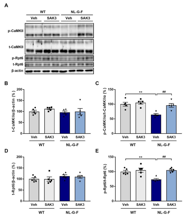
Figure 3.
SAK3 administration improves CaMKII-Rpt6 signaling in NL-G-F mice. (A) Representative image of western blot membranes containing hippocampal protein probed with antibodies against autophosphorylated CaMKII (T286), CaMKII, phosphorylated Rpt6 (S120), Rpt6, and β-actin. (B) Quantitative analyses of CaMKIIα, (C) autophosphorylated CaMKIIα (T286), (D) Rpt6, and (E) phosphorylated Rpt6 (S129) protein levels (n = 5 per group); autophosphorylated CaMKIIα: F (3, 16) = 11.01, p < 0.01; phosphorylated Rpt6: F (3, 16) = 9.316, p < 0.01. Error bars represent SEM. ** p < 0.01 vs. vehicle-treated WT mice; ## p < 0.01 vs. vehicle-treated NL-G-F mice. The symbols (solid dots, squares, triangles and inverted triangle) indicate the individual values.

