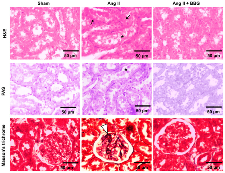
Figure 3.
Representative histological microphotographs stained with hematoxylin and eosin (H&E, first row), periodic acid Schiff (PAS, second row), and Masson´s trichrome (third row) in renal cortex of rats in Sham, Ang II and Ang II + BBG groups (n = 7 per group). In the Ang II group, there are areas of tubulointerstitial cell injury with intratubular debris indicated with an asterisk (*), focal areas of mononuclear infiltration indicated by black arrows and modest segmental mesangial widening in the glomeruli (white arrow head, ∇) are reduced in the Ang II + BBG group. Ang II = angiotensin II; BBG = brilliant blue G.

