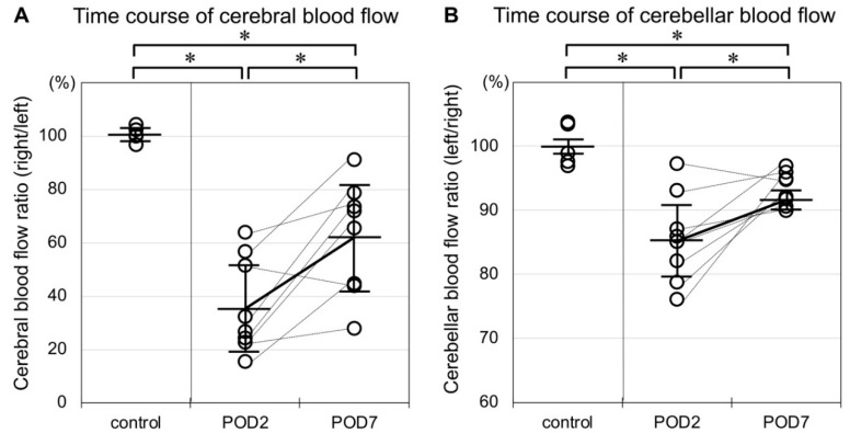
Figure 2.
(A) Rats in the control group showed no apparent laterality of cerebral blood flow. Cerebral blood flow ratio (right/left) was significantly decreased two and seven days after MCAO. The plots of each individual are connected with bottled lines. This ratio significantly recuperated over time. (B) Rats in the control group showed no apparent laterality of cerebellar blood flow. Cerebellar blood flow ratio (left/right) was significantly decreased two and seven days after MCAO, but this decrease was of a lesser degree than that in the cerebrum. As in the cerebrum, the decrease in blood flow ratio in the cerebellum likewise recuperated over time. (* p < 0.05, n = 5 in the control group, n = 8 in the MCAO group).

