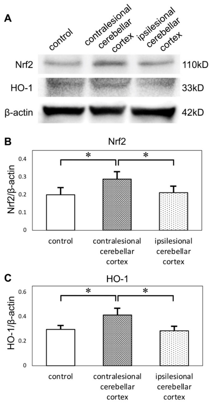
Figure 4.
(A) Western blots of cerebellar cortices show expression of Nrf2 and HO-1. Protein levels were normalized to beta-actin. (B) Quantification of western blots by densitometric analysis indicated that the expression of Nrf2 was upregulated in contralesional cerebellar cortices. (C) The expression of HO-1 was also upregulated in contralesional cerebellar cortices (* p < 0.05 versus other groups, n = 6 in each group). Nrf2: nuclear factor erythroid 2-related factor 2, HO-1: heme oxygenase-1.

