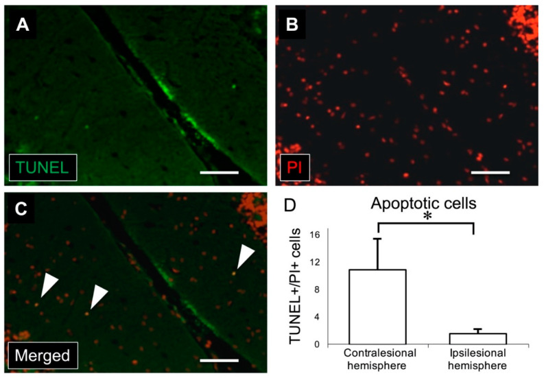
Figure 5.
(A) Immunostaining for TUNEL (green) shows apoptotic cells in the molecular layer of the cerebellar cortex. (B) Nuclei were stained with PI in red. (C) Merged image of TUNEL and PI immunostaining. White arrowheads indicate TUNEL/PI-positive cells. (D) There was a significant increase in the number of TUNEL/PI-positive cells in the contralesional (left) cerebellar cortex compared to the ipsilesional (right) cerebellar cortex (scale bar: 50 μm, * p < 0.001, n = 11 in each group). TUNEL: terminal deoxynucleotidyl transferase deoxyuridine triphosphate nick-end labeling, PI: propidium iodide.

