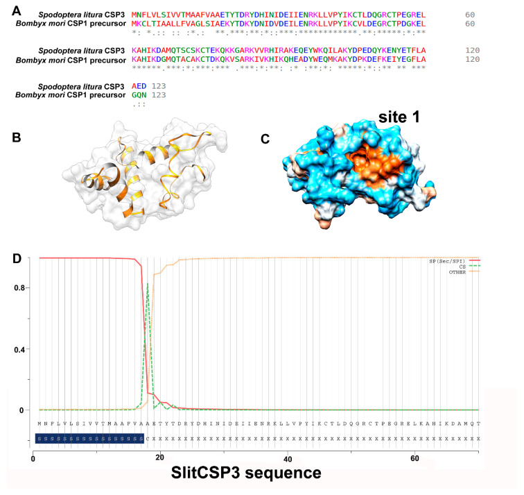
Figure 1.
(A) Sequence alignment to see the conservation between SlitCSP3 and B. mori CSP1 precursor sequence. The sequences are well-conserved except at the N-terminus, which was not included during structure modeling. (B) Three-dimensional model predicted through homology modeling. (C) A representation of its hydrophobic surface with orange depicting the hydrophobic core. (D) The prediction of signal peptide sequence present in SlitCSP3 at the N-terminus till 17th residue, and 18th is the cleavage site.

