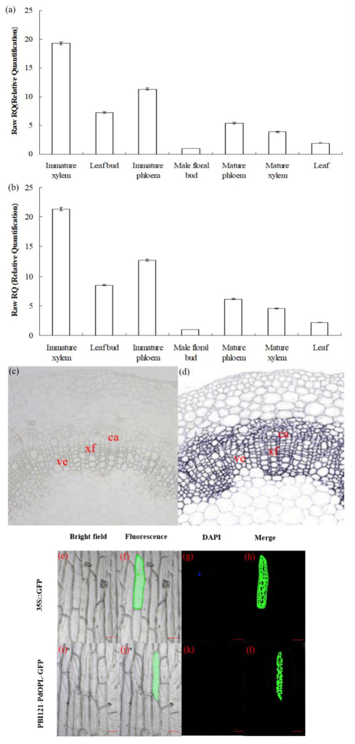
Figure 2.
Expression pattern and subcellular localization of the PdOLP1 protein. (a,b) PdOLP1 expression in various tissues was analyzed by quantitative real-time PCR (qRT-PCR). Expression was normalized to α-tubulin (TUA1) (a) and ubiquitin (UBQ1) reference genes (b). Error bars: ± standard error (SE) of three replicates. (c,d) In situ localization of PdOLP1 in P. deltoides stems. Sections of the stems were hybridized with digoxigenin-labeled antisense (c) or sense (d) probes. The hybridization signals are shown in purple. ve, vessel; xf, xylary fiber; ca, cambium. Scale bars = 25 µm. (e–l) Subcellular localization of PdOLP1. The green fluorescent signal of PBI121-PdOLP1-GFP was detected within the cytoplasm of onion epidermal cells (j,l), and green fluorescent protein (GFP) signal alone was localized in the cytoplasm, nucleus (4′,6-diamidino-2-phenylindole (DAPI) stained) and plasma membrane (f,h). Scale bars = 100 µm.

