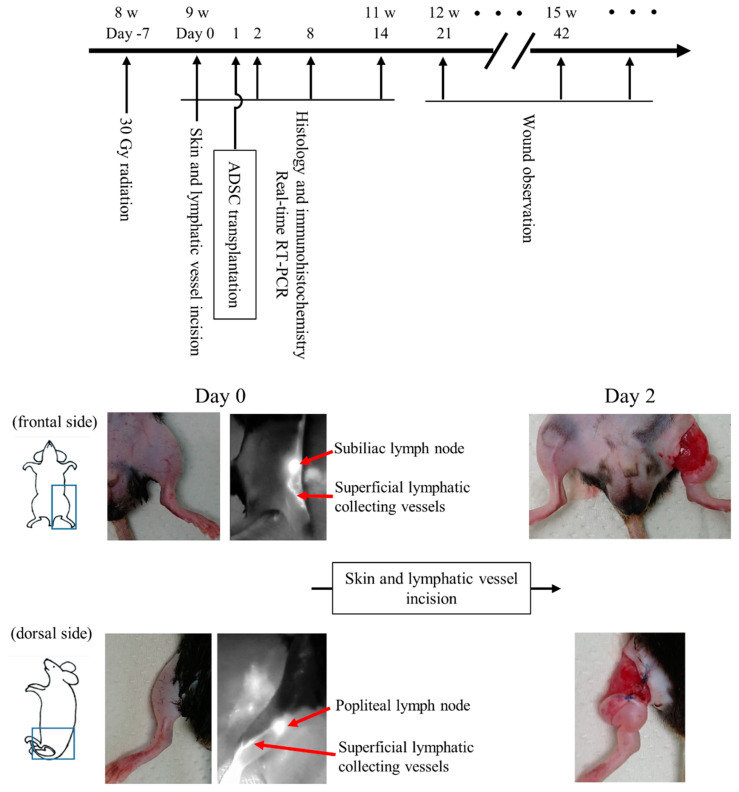
Figure 1.
Time course of the experiment and macroscopic image of mouse secondary lymphedema model. Experimental time course and images of skin and lymphatic vessel incisions. The incised lymphatic vessels were identified using a fluorescence near-infrared video camera with intradermal injection of indocyanine green.

