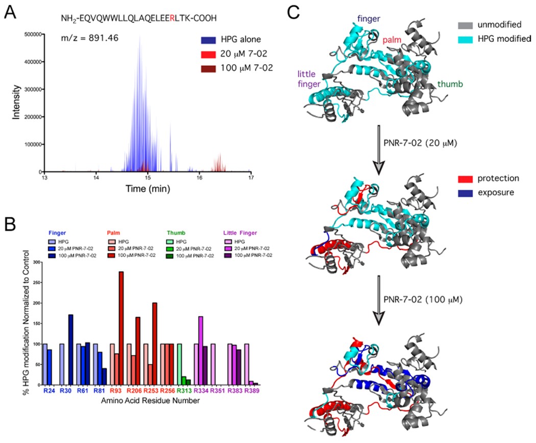
Figure 4.

Protein footprinting identifies the PNR-7-02 binding site on the hpol η little finger. (A) Selected ion chromatogram for the HPG-modified peptide containing Arg351 in the presence of HPG alone (blue), HPG + 20 μM PNR-7-02 (red), and 100 μM PNR-7-02 (dark red). (B) Relative abundance of HPG-modified arginine was quantified from MS data. The fraction of HPG-modified arginine observed in the presence of PNR-7-02 was normalized to that observed in the absence of the compound. The modified arginine residues in the finger, palm, thumb, and little finger domains of hpol η are shown in blue, red, green, and purple, respectively. (C) Cartoon representation of hpol η (PDB ID 3MR2) shown with regions of the protein containing HPG-modified residues in cyan and regions that were not modified by HPG in gray. The addition of PNR-7-02 caused changes in HPG reactivity for multiple arginines. Peptides with arginine residues that displayed a greater than 2-fold reduction in HPG reactivity (i.e., protection) are shown in red, whereas regions that exhibited a greater than 2-fold increase in HPG reactivity (i.e., exposure) are shown in blue.
