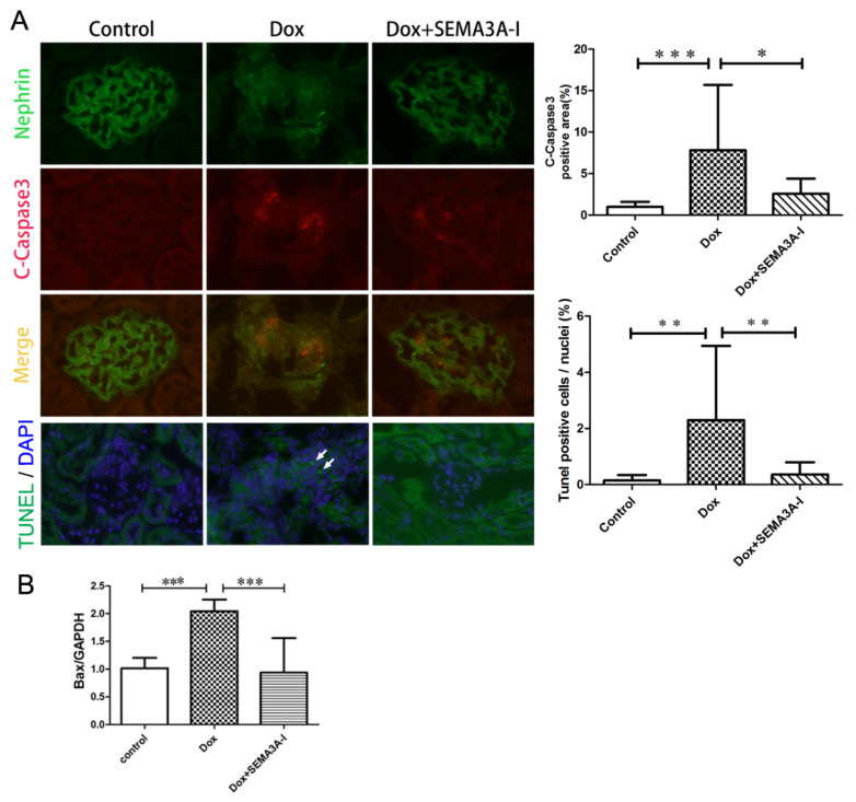
Figure 3.
SEMA3A-inhibitor protected from Doxorubicin-induced podocyte apoptosis. (A) Dual immunofluorescence staining of cleaved-caspase3 (C-Caspase3) (red) and nephrin (green) in mouse glomeruli from the control, Dox, and Dox + SEMA3A-I groups at the time point of 2 weeks after Dox injection, showing the increase of C-Caspase3-positive podocytes in the Dox group, and fewer C-Caspase3-positive cells in the Dox + SEMA3A-I group. Images of immunofluorescent staining of TdT-mediated dUTP Nick-End Labeling (TUNEL, green) and 4′,6′-diamidino-2-phenylindole (DAPI, blue) in the control, Dox, and Dox + SEMA3A-I groups (Lowest panel) show that TUNEL-positive cells were detected in the Dox group (white arrows), while almost no TUNEL-positive cells were detectable in the control and Dox + SEMA3A-I groups. Representative images are shown. Original magnification, ×400 (C-Caspase3 and nephrin) and x200 (TUNEL). C-Caspase3-positive area/glomeruli (%) and TUNEL-positive cells/nuclei (%) are shown in the graphs. (B) Reverse transcription-quantitative polymerase chain reaction (RT-qPCR) analysis of B cell lymphoma2-associated x-protein (Bax)/Glyceraldehyde 3-phosphate dehydrogenase (GAPDH) ratio in the control, Dox, and Dox + SEMA3A-I groups at the time point of 2 weeks after Dox injection, showing the increase of Bax mRNA level in the Dox group. * p < 0.05, ** p < 0.01, *** p < 0.001.

