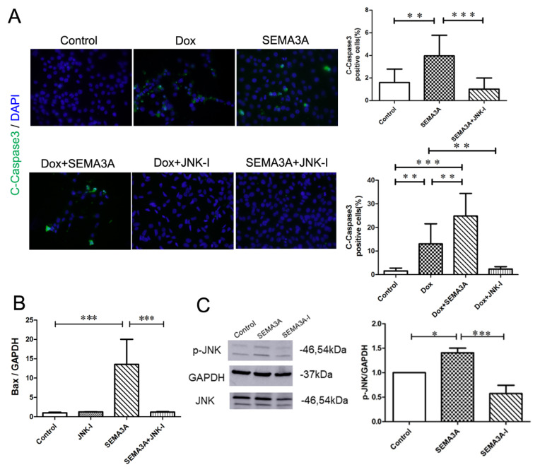
Figure 6.
JNK-inhibitor blocked SEMA3A-induced podocyte apoptosis. (A) Immunofluorescence staining of cleaved-caspase 3 (C-Caspase3) (green) and DAPI (blue) in podocytes with or without Dox (0.5 μg/mL), SEMA3A (50 ng/mL), Dox (0.5 μg/mL) + SEMA3A (50 ng/mL), Dox (0.5 μg/mL) + JNK-inhibitor (JNK-I) (10 μM), SEMA3A (50 ng/mL) + JNK-I (10 μM), showing that JNK-I prevented from Dox- and SEMA3A-induced podocyte apoptosis. Representative images are shown. Original magnification, ×200. C-Caspase3-positive cells/nuclei (%) in 200× fields are shown in the graphs. (B) RT-qPCR analysis of Bax and GAPDH mRNA with or without SEMA3A (50 ng/mL), JNK-I (10 μM), and SEMA3A (50 ng/mL) + JNK-I (10 μM), showing that mRNA level of Bax is increased with SEMA3A treatment, and is decreased with the JNK-I treatment. (C) Western blotting analysis of p-JNK, JNK, and GAPDH with or without SEMA3A (50 ng/mL) or SEMA3A-I (0.5 μM), showing that SEMA3A upregulated the expression of p-JNK. Densitometric analysis was performed to quantify the Western blotting results. Data are shown from three independent experiments. * p < 0.05, ** p <0.01, *** p < 0.001.

