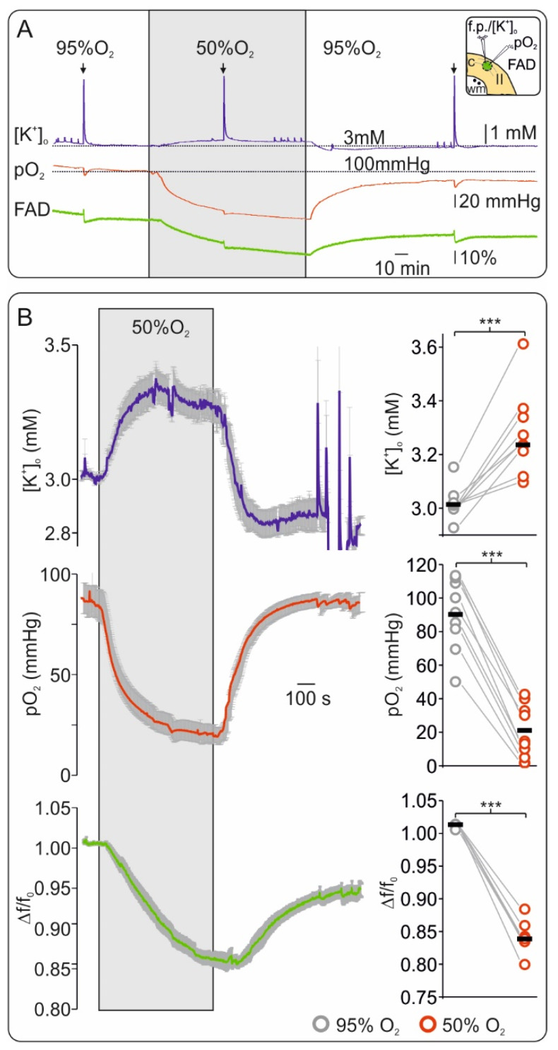
Figure 1.
Flavin adenine dinucleotide (FAD) fluorescence is highly sensitive to short hypoxia in vitro. (A) Exemplary experiment of simultaneous recording of extracellular potassium ([K+]o) (upper trace blue), tissue oxygenation (pO2) (middle trace, red) and FAD (bottom trace, green) showing the effects of short hypoxia (application of 50% oxygen, grey surface) and reoxygenation. Decreasing O2 supply generated a slight increase in interstitial K+ and a marked reductive shift of FAD fluorescence. Inflections in the traces resulted from electrical stimulation (arrows on top represent stimulation events of 20 Hz during 2 s, see Figure 2). Thus, the FAD signal was highly sensitive and timely coupled to changes in O2 supply. Right on top: graphical representation of cortical slice detail and recording technic (c: neocortex, wm: white matter, II: layer II). Ion sensitive electrode and O2 electrode were positioned in layer II of the frontal cortex in the area of interest for FAD imaging (green circle) while electrical stimulation was performed in the adjacent white matter (black dots). (B) Left: Average curves (with standard errors) for all experiments concerning changes in [K+]o (top, blue line), pO2 (middle, red line) and FAD (bottom, green line). Right: statistic plots of single values for each experiment (grey circles: control, red circles: hypoxia, black lines: median values). All parameters were recorded simultaneously, n = 9, *** = p < 0.001.

