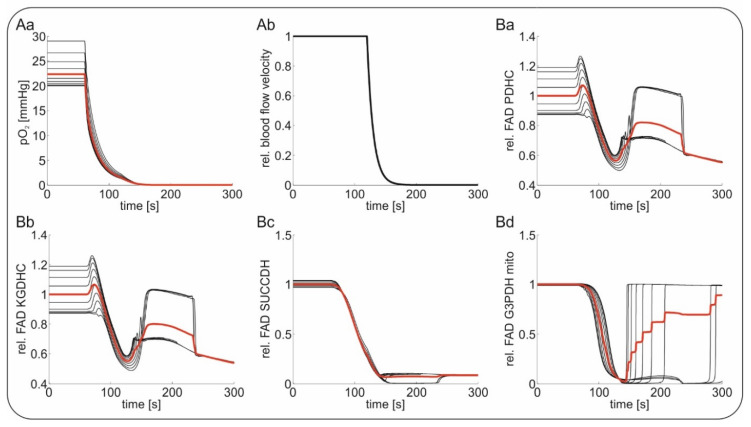
Figure 6.
Stimulation of in vivo Flavin adenine dinucleotide (FAD) fluorescence during hypoxia and subsequent global ischemia-induced terminal spreading depolarization (SD); capillary tissue oxygenation (pO2) was decreased from 30 mmHg to 0 mmHg at t = 60 s followed by further substrate depletion during terminal SD. Red traces indicate average values of the black traces indicating the different layers around the vessel. (Aa) The course of the decrease in oxygen during simulated hypoxia mimicking in vivo experiments; (Ab) followed by maintained hypoxia, the relative blood flow velocity in the tissue was stopped, simulating the events of global ischemia and terminal SD; simulated relative changes signal of FAD bound to pyruvate dehydrogenase (PDHC, (Ba)); to α-ketogluterate dehydrogenase (KGDHC, (Bb)); to succinate dehydrogenase (SUCCDH, (Bc)) and to mitochondrial glycereol-3-phophate dehydrogenase (G3PDH, (Bd)).

