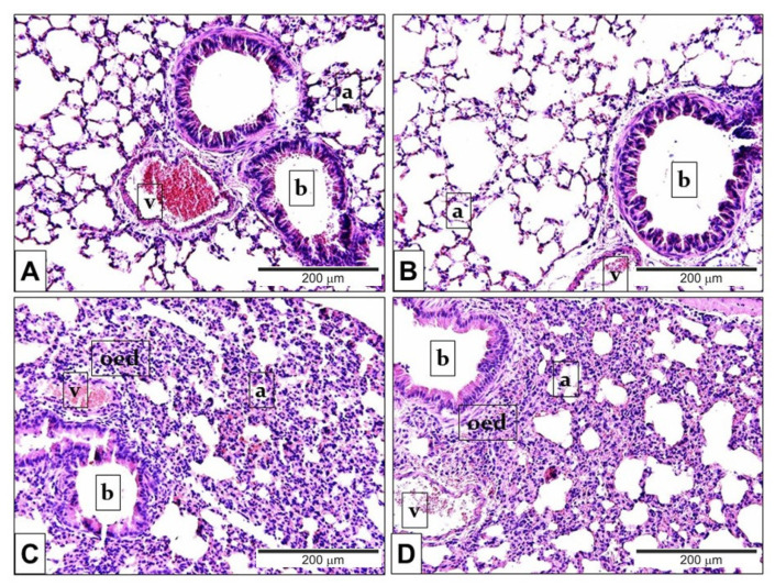
Figure 3.
Representative histological pictures of LPS-induced pulmonary alterations. Compared to the PBS-treated (A) Trpa1+/+ and (B) Trpa1−/− control groups, the lung tissue of LPS-treated (C) Trpa1+/+ and (D) Trpa1−/− mice exhibited remarkable perivascular and peribronchial oedema, neutrophil granulocyte accumulation, macrophage infiltration and goblet cell hyperplasia (hematoxylin-eosin staining; 200× g magnification; a: alveolus, b: bronchiolus, v: venula, oed: oedema).

