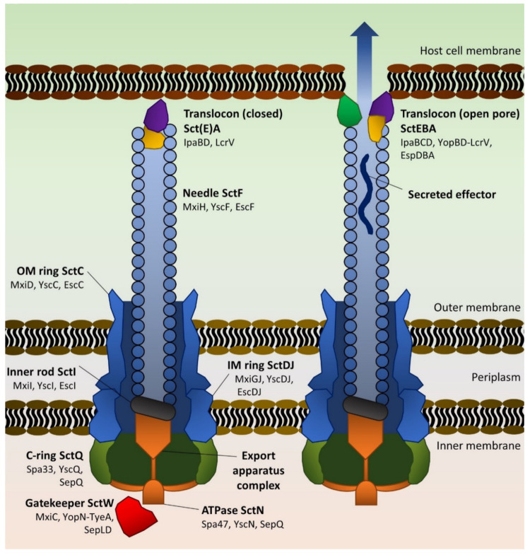
Figure 1.
Schematic of type 3 secretion systems. The major structural rings (C-ring in olive, inner/outer membrane scaffold rings in blue) support the ATPase-containing export apparatus (orange), which is linked via an inner rod adaptor helix to the needle filament (grey oblongs and blue circles, respectively). Tip and gatekeeper proteins (purple, yellow, red) initially block the needle and prevent effector translocation (left) until the complex senses host cell contact—note that SctE (IpaB) has only been shown to play a role in blocking secretion in Shigella. Rearrangements then permit hydrophobic pore formation (purple, green) in the eukaryotic membrane and effector secretion (right). Proteins are not shown to scale. Adapted from [28,29,30,31].

