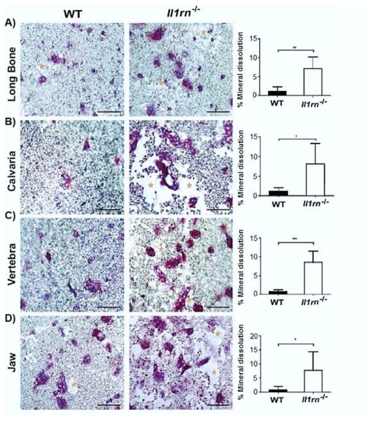
Figure 5.
Il1rn−/− osteoclasts show an enhanced mineral dissolution independently of the skeletal site. Absence of IL-1RA increased the in vitro dissolution of the inorganic matrix of bone from all osteoclast precursors irrespective of the isolation site, suggesting that site-specific differences present in vivo can be outweighed in vitro. (A–D) Microphotographs of hydroxyapatite-coated plates of which part is dissolved by osteoclasts derived from various skeletal sites in both wild-type (WT) and Il1rn−/− mice. Bone marrow (BM) cells from long bone, calvaria, vertebra, and jaw were cultured with 30 ng/mL M-CSF and 20 ng/mL RANKL on hydroxyapatite-coated plates for 8 days. Osteoclasts were stained by TRAcP (in purple) and nuclei were stained by 4’,6-Diamidino-2-Phenylindole (DAPI) (blue). The dissolved area was labeled by asterisks. Scale bar = 100 µm. The dissolved area was quantified and compared between WT and Il1rn−/− mice for long bone (A), calvaria (B), vertebra (C) and jaw (D) (n = 6, *p < 0.05, **p < 0.01, ***p < 0.001).

