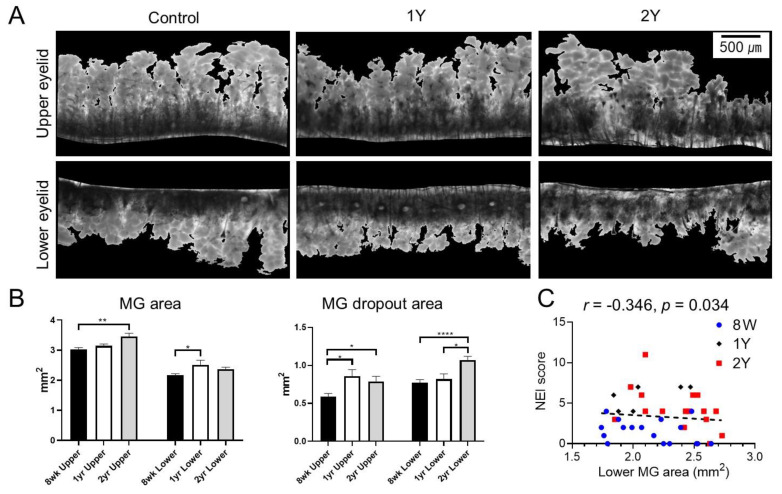
Figure 2.
(A) Representative transillumination meibography images. (B) Meibomian gland (MG) areas of the upper eyelid in 2Y-old mice (p = 0.004; Kruskal–Wallis test followed by Dunn’s post hoc test) and lower eyelid in 1Y-old mice (p = 0.021; one-way ANOVA followed by Tukey’s post hoc test) were larger than those in 8W-old mice. MG dropout areas of upper eyelids were larger in 1Y- and 2Y-old mice than 8W-old mice (p = 0.023 and p = 0.02, respectively; Kruskal–Wallis test followed by Dunn’s post hoc test). MG dropout areas of lower eyelids were larger in 2Y-old mice than 8W- and 1Y-old mice (p < 0.0001 and p = 0.029, respectively; one-way ANOVA followed by Tukey’s post hoc test). (C) The age-adjusted MG area of the lower eyelid was negatively correlated with the National Eye Institute (NEI) score (r = −0.346; p = 0.034; partial correlation analysis). * p < 0.05, ** p < 0.01, and **** p < 0.0001. Data are presented as means ± standard error.

