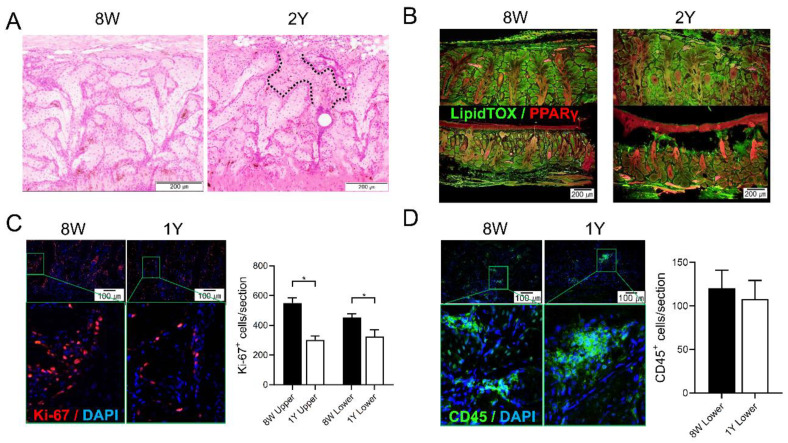
Figure 3.
(A) Representative images of coronal sections of the MGs stained with hematoxylin-eosin (H&E). The area between the dotted lines indicates suspicious MG dropout (200×). (B) Representative images of coronal sections of the MGs stained with LipidTOX and PPARγ immunofluorescence (100×). (C) The expression of Ki67+ cells in both MGs of the upper and lower eyelids was significantly reduced in 1Y-old mice compared to 8W-old mice (p = 0.036 by the Mann–Whitney test and 0.035 by the independent t-test, respectively; 200×). (D) The expression of CD45+ cells in the MGs of the lower eyelid was not different between 8W- and 1Y-old mice (p = 0.571; Mann–Whitney test; 200×). All images were obtained from the coronal section. n = 5 and 3 for 8W- and 1Y-old mice, respectively. * p < 0.05.

