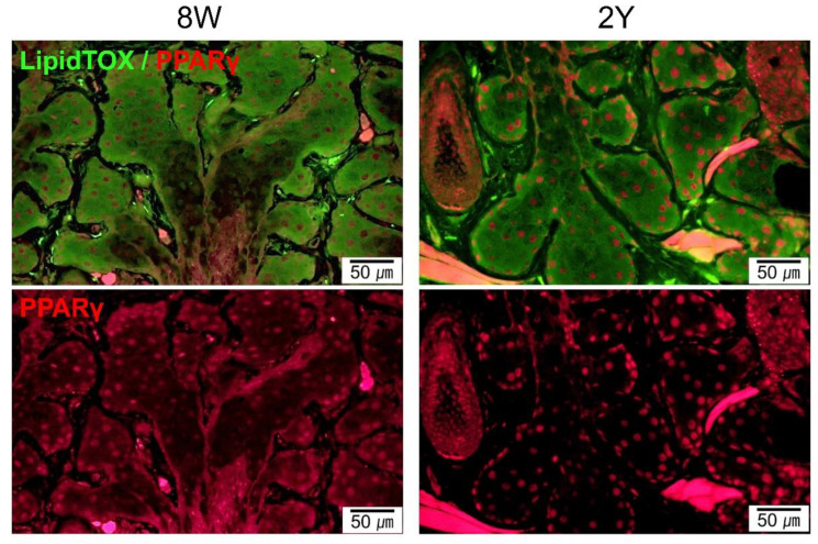
Figure 4.
Representative images of coronal sections of the MGs stained with LipidTOX and PPARγ immunofluorescence (coronal section; 400×). Lipid droplets showed a similar distribution for 8W- and 2Y-old mice. PPARγ staining shows cytoplasmic and nuclear localization in 8W-old mice. In contrast, PPARγ staining shows predominant nuclear localization in 2Y-old mice.

