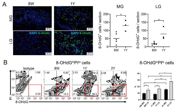
Figure 7.
(A) Immunofluorescence staining of 8-OHdG in the MGs (sagittal section, upper panel) and LGs (lower panel; 400×). The number of 8-OHdG+ cells was significantly higher in 1Y-old mice (n = 5) than in 8W-old mice (n = 3) in both the MGs and LGs (p = 0.018 and 0.015, respectively; independent t-test). (B) The percentages of 8-OHdGhi PIlo cells were significantly higher in the LGs than in the MGs in 8W- and 1Y-old mice (p = 0.024 and 0.004, respectively; independent t-test). In both the MGs and LGs, 8-OHdGhi PIlo cells were significantly higher in 2Y-old mice than in 8W- and 1Y-old mice (one-way ANOVA; n = 9, 5, and 3 for 8W-, 1Y-, and 2Y-old mice, respectively). Red boxes indicate 8-OHdGhi PIlo cells. * p < 0.05 and ** p < 0.01. Data are presented as means ± standard error.

