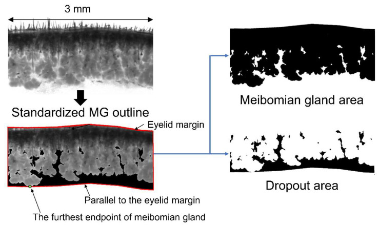
Figure 9.
Ex vivo transillumination meibography images of the central 3-mm area of interest were analyzed. “The standardized MG outline” was defined with an imaginary line starting at the furthest endpoint of the MG and parallel to the eyelid margin. The area of the MG, including the eyelid margin, was defined as “the MG area”, and the area where the MG does not exist inside the standardized MG outline was defined as “the dropout area”.

