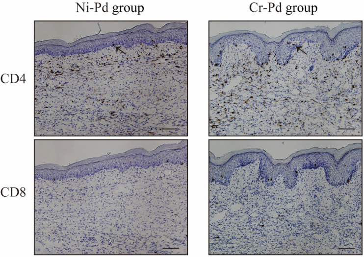
Figure 4.
Immunohistochemical (IHC) analyses of CD4 and CD8 in the mouse model of cross-reactive metal allergy. CD3+ T cells infiltrated into the inflamed skin in the Ni-Pd and Cr-Pd groups. CD4+CD8− T cells were present in the epithelial basal layer and the upper dermis (arrows). Scale bar = 10 µm.

