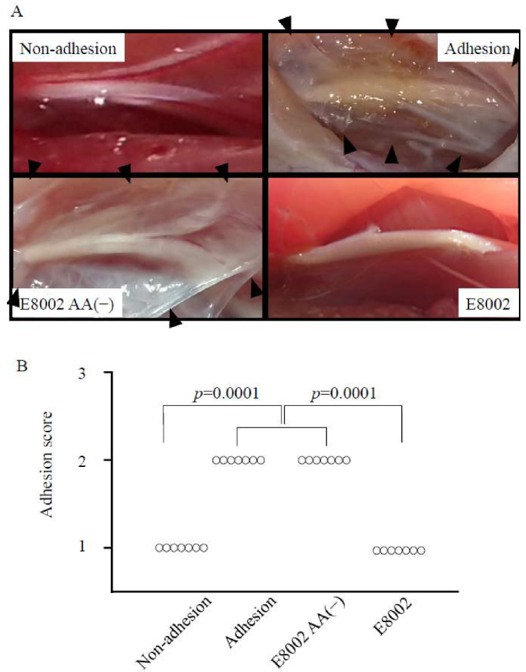
Figure 3.

Effect of E8002 membrane on macroscopic peripheral nerve adhesions at 6 weeks post-surgery. (A) Representative photomicrographs of the macroscopic appearance of the adhesion site (adhesion, E8002(−), and E8002 groups) or related area (non-adhesion group). The tissue located in the center of each figure is the nerve tissue. The adhesion area is recognizable as fibrous connective tissue located around the nerve tissue (the white area indicated by black arrowheads). (B) Adhesion scores in the non-adhesion group and the E8002 group were significantly lower than those in the adhesion group and the E8002 l-ascorbic acid (AA(−)) group (n = 7 adhesion sites or related areas per group). p = 0.001 as determined by the Kruskal–Wallis test.
