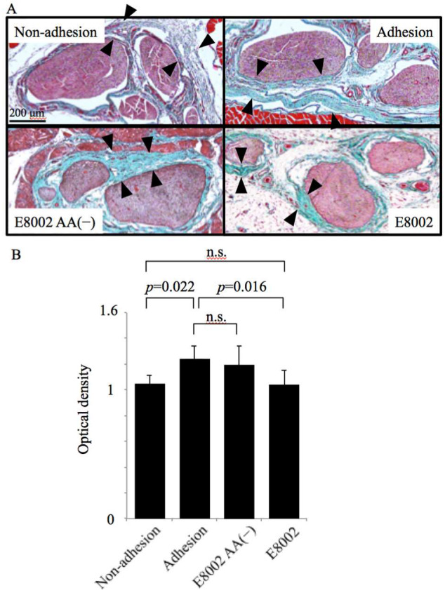
Figure 4.

Effect of the E8002 membrane on microscopic appearance of peripheral nerve adhesions at 6 weeks post-surgery. (A) Representative photomicrographs of the sciatic nerve and neural bed stained with aldehyde fuchsin Masson–Goldner staining (scale bar, 200 µm). Pink-stained nerve tissue located in the center is surrounded by connective tissue (light blue/green area indicated by black arrowheads). In the E8002 group, there was thin and loose connective tissue around the nerve, with space (no staining) surrounding the connective tissue. This space was considered to be a trace of E8002 absorption. (B) Quantitative analysis of the optical density of the Masson–Goldner staining (n = 7 adhesion sites or related areas per group). Values are mean ± SE; n.s., non-significant determined by one-way analysis of variance followed by the Bonferroni–Dunn correction.
